COMPARACIÓN MORFOLÓGICA DE CÉLULAS AISLADAS DE LA PULPA DENTAL OBTENIDAS DE TERCEROS MOLARES DE UN SOLO DONADOR
Resumen
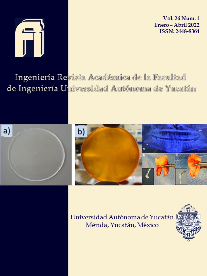
Actualmente, el estudio de las células troncales (CT) derivadas de la cavidad oral es de gran importancia debido a su capacidad de originar diferentes tipos celulares, por lo que podrían considerarse como una alternativa para el establecimiento de modelos de investigación in vitro. Particularmente, las células derivadas de la pulpa dental presentan una morfología fibroblastoide y una alta capacidad proliferativa para su diferenciación hacia múltiples linajes, lo cual las convierte en una alternativa viable para la implementación de cultivos celulares. Una de las características poco estudiadas para el establecimiento de los cultivos celulares de la pulpa dental, es el efecto de la posición del órgano dentario de donde se obtiene la pulpa para la generación de los explantes; por lo que en este estudio se comparó la proliferación celular de los terceros molares superior e inferior de un mismo paciente, observando que la proliferación celular es influenciada por la posición del diente y el tipo de raíz de éste.
Citas
Alraies, A., Alaidaroos, N. Y., Waddington, R. J., Moseley, R. y Sloan, A. J. (2017). Variation in human dental pulp stem cell ageing profiles reflect contrasting proliferative and regenerative capabilities. BMC Cell Biology 18(12). https://doi.org/10.1186/s12860-017-0128-x
Bressan, E., Ferroni, L., Gardin, C., Pinton, P., Stellini, E., Botticelli, D.,... & Zavan, B. (2012). Donor age-related biological properties of human dental pulp stem cells change in nanostructured scaffolds. PloS one, 7(11), e49146. https://journals.plos.org/plosone/article?id=10.1371/journal.pone.0049146
de Carvalho, R. W. F., de Araújo Filho, R. C. A., & do Egito Vasconcelos, B. C. (2013). Assessment of factors associated with surgical difficulty during removal of impacted maxillary third molars. Journal of Oral and Maxillofacial Surgery. 71(5), 839-845. https://pubmed.ncbi.nlm.nih.gov/23598549/
Dowthwaite, G. P., Bishop, J. C., Redman, S. N., et al. (2004). The surface of articular cartilage contains a progenitor cell population. Journal of Cell Science, 117(6), 889–897. https://pubmed.ncbi.nlm.nih.gov/14762107/
Fuentes, R., Borie, E., Bustos, L., y Thomas, D. (2009). Morfometría de terceros molares: un estudio de 55 casos. International Journal of Morphology, 27(4), 1285-1289. http://dx.doi.org/10.4067/S0717-95022009000400050
Gage, F. H. (2000). Mammalian neural stem cells. Science, 287 (5457), 1433–1438. https://pubmed.ncbi.nlm.nih.gov/10688783/
Gronthos, S., Brahim, J., Li, W. et al. (2002). Stem cell properties of human dental pulp stem cells. Journal of Dental Research, 81(8), 531–535. https://pubmed.ncbi.nlm.nih.gov/12147742/
Guerrero-Jiménez M, Nic-Can GI, Castro-Linares N, Aguilar-Ayala FJ, Canul-Chan M, Rojas-Herrera RA, Peñaloza-Cuevas R, Rodas-Junco BA. 2019. In vitro histomorphometric comparison of dental pulp tissue in different teeth. Peer J 7:e8212. https://doi.org/10.7717/peerj.8212
Huang G., Gronthos, S. y Shi, S. (2009). Mesenchymal stem cells derived from dental tissues vs those from other sources: their biology and role in regenerative medicine. Journal of Dental Research, 88(9) 792–806. https://www.ncbi.nlm.nih.gov/pmc/articles/PMC2830488/
Huard, J., Cao, B. y Qu-Petersen, Z. (2003). Muscle-derived stem cells: potential for muscle regeneration. Birth Defect Research, 69(3), 230-237. https://pubmed.ncbi.nlm.nih.gov/14671776/
Jones, R. J. y Matsui, W. (2007). Cancer stem cells: from bench to bedside. Biology of blood and marrow. Transplantation, 13, 47–52. https://pubmed.ncbi.nlm.nih.gov/18167509/
Kawashima, N. (2012). Characterisation of dental pulp stem cells: a new horizon for tissue regeneration? Archives of Oral Biology, 57, 1439–1458. https://pubmed.ncbi.nlm.nih.gov/22981360/
Kobayashi, T., Torii, D., Iwata, T., Izumi, Y., Nasu, M., y Tsutsui, T. W. (2020). Characterization of proliferation, differentiation potential, and gene expression among clonal cultures of human dental pulp cells. Human cell 33(3), 490. https://doi.org/10.1007/s13577-020-00327-9
Liu, H., Gronthos, S. y Shi, S. (2006). Dental pulp stem cells. Methods in Enzymology, 419, 99–113. https://pubmed.ncbi.nlm.nih.gov/17141053/
Longoni, A., Utomo, L., van Hooijdonk, I. E., Bittermann, G., Vetter, V. C., Spanjer, E. K.,.. y Gawlitta, D. (2020). The chondrogenic differentiation potential of dental pulp stem cells. Eur. Cells Mater 39, 121-135. https://doi.org/10.22203/ecm.v039a08
Lübbers, H. T., Matthews, F., Damerau, G., Kruse, A. L., Obwegeser, J. A., Grätz, K. W., y Eyrich, G. K. (2011). Anatomy of impacted lower third molars evaluated by computerized tomography: is there an indication for 3-dimensional imaging? Oral Surgery, Oral Medicine, Oral Pathology, Oral Radiology, and Endodontology, 111(5), 547-550. https://pubmed.ncbi.nlm.nih.gov/20952229
Mercado-Rubio, M. D., Pérez-Argueta, E., Zepeda-Pedreguera, A., Aguilar-Ayala, F. J., Peñaloza-Cuevas, R., Kú-González, A., Nic-Can, G. I. (2021). Similar features, different behaviors: A comparative in vitro study of the adipogenic potential of stem cells from human follicle, dental pulp, and periodontal ligament. Journal of Personalized Medicine, 11(8), 738. https://www.mdpi.com/2075-4426/11/8/738
Noth U, Osyczka AM, Tuli R, Hickok NJ, Danielson KG, Tuan RS (2002). Multilineage mesenchymal differentiation potential of human trabecular bone-derived cells. Journal Orthopaedic Research, 20(5), 1060–9. https://pubmed.ncbi.nlm.nih.gov/12382974/
Olguín Martínez, T. G., y Amarillas Escobar, E. D. (2017). Morfología radicular de los terceros molares. Revista ADM, 74(1). https://www.medigraphic.com/cgi-bin/new/resumen.cgi?IDARTICULO=70658
Patil R, Kumar BM, Lee WJ, et al. (2014). Multilineage potential and proteomic profiling of human dental stem cells derived from a single donor. Experimental Cell Research, 320(1), 92–107. https://europepmc.org/article/med/24162002
Rivas-Aguayo, A. G. (2018). Establecimiento de metodologías para el aislamiento y cultivo in vitro de células troncales de la pulpa dental humana de terceros molares. Tesis de Licenciatura de Ing. En Biotecnología. FIQ-UADY. Mérida pp 1–85
Rodas-Junco, B. A., Canul-Chan, M., Rojas-Herrera, R. A. et al. (2017). Stem cells from dental pulp: what epigenetics can do with your tooth. Frontiers in Physiology, 8(999), 1–20. https://pubmed.ncbi.nlm.nih.gov/29270128/
Rodríguez-Lozano, F. J., Insausti, C. L., Iniesta, F. et al. (2012). Mesenchymal dental stem cells in regenerative dentistry. Medicina oral, patologia oral y cirugia bucal, 17(6), 1062–1067. https://dialnet.unirioja.es/servlet/articulo?codigo=4499431
Ullah, I., Subbarao, R. B., Kim, E. J. et al. (2016). In vitro comparative analysis of human dental stem cells from a single donor and its neuronal differentiation potential evaluated by electrophysiology. Life Sciences, 154, 39–51. https://www.sciencedirect.com/science/article/pii/S0024320516302466
Vazquez, D., Hetch, P., & Martínez, M. E. (2012). Radicular synostosis: frequency study using panoramic x-rays as diagnostic method. Revista Odontológica Mexicana, 16(2). http://revistas.unam.mx/index.php/rom/article/view/30916

Esta obra está bajo licencia internacional Creative Commons Reconocimiento-NoComercial 4.0.
Avisos de derechos de autor propuestos por Creative Commons
1. Política propuesta para revistas que ofrecen acceso abierto
Aquellos autores/as que tengan publicaciones con esta revista, aceptan los términos siguientes:
- Los autores/as conservarán sus derechos de autor y garantizarán a la revista el derecho de primera publicación de su obra, el cuál estará simultáneamente sujeto a la Licencia de reconocimiento de Creative Commons que permite a terceros compartir la obra siempre que se indique su autor y su primera publicación esta revista.
- Los autores/as podrán adoptar otros acuerdos de licencia no exclusiva de distribución de la versión de la obra publicada (p. ej.: depositarla en un archivo telemático institucional o publicarla en un volumen monográfico) siempre que se indique la publicación inicial en esta revista.
- Se permite y recomienda a los autores/as difundir su obra a través de Internet (p. ej.: en archivos telemáticos institucionales o en su página web) antes y durante el proceso de envío, lo cual puede producir intercambios interesantes y aumentar las citas de la obra publicada. (Véase El efecto del acceso abierto).
